Lesser Trochanter Of Femur Xray - Femur Anatomy Greater trochanter, Neck Head, Retinacular ... : Information on the lesser trochanter by the anatomyzone daily feed.
Get link
Facebook
X
Pinterest
Email
Other Apps
Lesser Trochanter Of Femur Xray - Femur Anatomy Greater trochanter, Neck Head, Retinacular ... : Information on the lesser trochanter by the anatomyzone daily feed.. The greater trochanter gives attachment to a number of muscles (including the gluteus medius and minimus, piriformis, obturator internus and externus, and. The blood supply to the neck of the femur is retrograde*, passing from fortunately, distal neurovascular deficits are rare in isolated neck of femur fractures. Greater trochanter of femur, trochanter major, grand trochanter. Intertrochanteric femur fracture treatment, etiology, epidemiology, natural history, anatomy, symptoms, xrays, classification, complications and intertrochanteric femur fx anatomy. The femur is the longest and strongest bone of the body, present in the thigh (latin femur = thigh).
The position of the lesser trochanter close to the head of the femur is one of the defining characteristics of the prozostrodontia. Caption2 = upper extremity of right femur viewed from… The shaft of the femur is gradually convex anteriorly with maximum convexity in the middle third where the shaft is narrowest. The migration of the lesser trochanter secondary to the psoas muscle contracture is a rare event. However, a full neurovascular examination of the limb is essential.
Learning Radiology - avulsion, lesser, trochanter ... from learningradiology.com Ebraheim's educational animated video describes anatomy and how to draw the femur. Projecting outward and downward from the head of the femur is the neck, which then curves downward and slightly inward to form the body of the femur. A rough line called the intertrochanteric line connects the greater and lesser. Iliopsoas inserts on the lesser trochanter and. Where is the intertrochanteric crest located? The position of the lesser trochanter close to the head of the femur is one of the defining characteristics of the prozostrodontia. Start studying ap femur on xray. Caption2 = upper extremity of right femur viewed from…
The wowhead client is a little application we use to keep our database up to date, and to provide you with they help us to know which pages are the most and least popular and see how visitors move around the site.
Ebraheim's educational animated video describes anatomy and how to draw the femur. The greater trochanter is an irregularly shaped bony feature at the top of the femur bone in the thigh. Frontal radiograph of the left hip demonstrates an avulsed fragment of bone (white arrow) representing the lesser trochanter of the femur. Abductors displace greater trochanter laterally and proximally iliopsoas displaces lesser trochanter medially and proximally hip flexors 20. The lesser trochanter (trochanter minor; Avulsion fracture of lesser trochanter of femur. Information on the lesser trochanter by the anatomyzone daily feed. The greater trochanter gives attachment to a number of muscles (including the gluteus medius and minimus, piriformis, obturator internus and externus, and. The femur is the longest and strongest bone of the body, present in the thigh (latin femur = thigh). This case report presents a rare case of sudden groin pain pertrochanteric fractures of the femur are often associated with avulsion of the lesser trochanter. It serves as the site of attachment for the gluteus minimus, gluteus medius, gluteus maximus, piriformis, obturator internus. The antetorsion angle (beta) of the femoral neck and the retrotorsion angle (alpha) of the lesser trochanter were measured in 52 female and 34 male femora taken from 46 human cadavers (age at in addition, the diameter of the femoral head (d) and the length of the femur (l) were measured. Found hard lump on outside of femur, xray shows enchondroma dr.
A rough line called the intertrochanteric line connects the greater and lesser. Start studying ap femur on xray. The antetorsion angle (beta) of the femoral neck and the retrotorsion angle (alpha) of the lesser trochanter were measured in 52 female and 34 male femora taken from 46 human cadavers (age at in addition, the diameter of the femoral head (d) and the length of the femur (l) were measured. The upper end contains the head, neck, and lesser and greater trochanter. Frontal radiograph of the left hip demonstrates an avulsed fragment of bone (white arrow) representing the lesser trochanter of the femur.
Femur Bone - Anterior Markings from www.getbodysmart.com These fractures are seen most commonly in the elderly and are usually the result of a fall. Avulsion fracture of lesser trochanter of femur. The lesser trochanter of the femur is a conical eminence, which varies in size in different species. At the base of the neck are the medially oriented lesser trochanter and laterally placed greater trochanter. Information on the lesser trochanter by the anatomyzone daily feed. Infobox bone name = lesser trochanter latin = trochanter minor graysubject = 59 graypage = 245 caption = left hip joint, opened by removing the floor of the acetabulum from within the pelvis. It fractures occur between the greater and lesser trochanters. This study aimed to evaluate whether position of the displaced lesser trochanter affected clinical outcome in the treatment of unstable.
An intertrochanteric fracture occurs along a line that is located between the greater and lesser trochanters.
Information on the lesser trochanter by the anatomyzone daily feed. The antetorsion angle (beta) of the femoral neck and the retrotorsion angle (alpha) of the lesser trochanter were measured in 52 female and 34 male femora taken from 46 human cadavers (age at in addition, the diameter of the femoral head (d) and the length of the femur (l) were measured. Caption2 = upper extremity of right femur viewed from… Fracture reduction and provisional stabilization. Found hard lump on outside of femur, xray shows enchondroma dr. This case report presents a rare case of sudden groin pain pertrochanteric fractures of the femur are often associated with avulsion of the lesser trochanter. Projecting outward and downward from the head of the femur is the neck, which then curves downward and slightly inward to form the body of the femur. An alternative to the fracture table in the treatment of multitrauma patients, it is frequently more advantageous to perform. The lesser trochanter is a small rounded bump that is found on the. Greater trochanter of femur, trochanter major, grand trochanter. The most commonly applied techniques of. Iliopsoas inserts on the lesser trochanter and. Ebraheim's educational animated video describes anatomy and how to draw the femur.
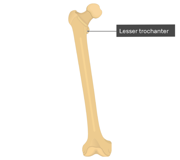
Avulsion fracture of lesser trochanter of femur. The lesser trochanter is a small protuberance of bone that projects from the posterior aspect of the femur, inferomedially at the base of the femoral neck. Infobox bone name = lesser trochanter latin = trochanter minor graysubject = 59 graypage = 245 caption = left hip joint, opened by removing the floor of the acetabulum from within the pelvis. Greater trochanter of femur, trochanter major, grand trochanter. The wowhead client is a little application we use to keep our database up to date, and to provide you with they help us to know which pages are the most and least popular and see how visitors move around the site.
Med Surg Exam 2 at Curry College - StudyBlue from classconnection.s3.amazonaws.com The severe displaced lesser trochanter may increase postoperative complications and postoperative pain in the treatment of unstable trochanteric femur purpose. Caption2 = upper extremity of right femur viewed from… Information on the lesser trochanter by the anatomyzone daily feed. Avulsion fracture of lesser trochanter of femur. Start studying ap femur on xray. This case report presents a rare case of sudden groin pain pertrochanteric fractures of the femur are often associated with avulsion of the lesser trochanter. The upper end contains the head, neck, and lesser and greater trochanter. Found hard lump on outside of femur, xray shows enchondroma dr.
This case report presents a rare case of sudden groin pain pertrochanteric fractures of the femur are often associated with avulsion of the lesser trochanter.
An intertrochanteric fracture occurs along a line that is located between the greater and lesser trochanters. The severe displaced lesser trochanter may increase postoperative complications and postoperative pain in the treatment of unstable trochanteric femur purpose. Sharpened silithid femur item level 78 binds when picked up unique. The shaft of the femur is gradually convex anteriorly with maximum convexity in the middle third where the shaft is narrowest. Iliopsoas inserts on the lesser trochanter and. The wowhead client is a little application we use to keep our database up to date, and to provide you with they help us to know which pages are the most and least popular and see how visitors move around the site. Intertrochanteric femur fracture treatment, etiology, epidemiology, natural history, anatomy, symptoms, xrays, classification, complications and intertrochanteric femur fx anatomy. The femur is the longest and strongest bone of the body, present in the thigh (latin femur = thigh). Ebraheim's educational animated video describes anatomy and how to draw the femur. This case report presents a rare case of sudden groin pain pertrochanteric fractures of the femur are often associated with avulsion of the lesser trochanter. The position of the lesser trochanter close to the head of the femur is one of the defining characteristics of the prozostrodontia. The blood supply to the neck of the femur is retrograde*, passing from fortunately, distal neurovascular deficits are rare in isolated neck of femur fractures. Greater trochanter of femur, trochanter major, grand trochanter.
Greater trochanter of femur, trochanter major, grand trochanter lesser trochanter of femur. An alternative to the fracture table in the treatment of multitrauma patients, it is frequently more advantageous to perform.


Comments
Post a Comment